PNAS:揭示肺粘液阻止纳米颗粒进入肺部
2013-05-06 ZinFingerNase 生物谷
2012年10月26日--来自德国萨尔布吕肯大学(Saarland University)和亥姆霍兹传染病研究中心(Helmholtz Centre for Infection Research, HZI)的研究人员阐明了肺粘膜的物理性质:他们发现肺粘膜上的刚性凝胶支架(rigid gel scaffold)将大的充满液体的孔隔离开,并且阻止纳米颗粒在单个孔的界限外运动。他们的发现加深了我们对呼吸

2012年10月26日--来自德国萨尔布吕肯大学(Saarland University)和亥姆霍兹传染病研究中心(Helmholtz Centre for Infection Research, HZI)的研究人员阐明了肺粘膜的物理性质:他们发现肺粘膜上的刚性凝胶支架(rigid gel scaffold)将大的充满液体的孔隔离开,并且阻止纳米颗粒在单个孔的界限外运动。他们的发现加深了我们对呼吸系统疾病(特别是传染性疾病)的理解,从而有助于开发新的吸入性药物。相关研究结果发表在PNAS期刊上。
粘膜包被着我们呼吸道的内表面。粘性的凝胶让肺部保持潮湿,并阻止病毒和其他的小颗粒如柴油机炭黑颗粒不受制止地进入体内。在此之前,科学家们仍不清楚这些纳米颗粒多大程度上能够穿过肺粘膜。在当前的这项研究中,研究人员获得的研究证据反驳了这一点。
论文通信作者Claus-Michael Lehr教授解释道,“肺粘膜是一种非常特别的凝胶。”
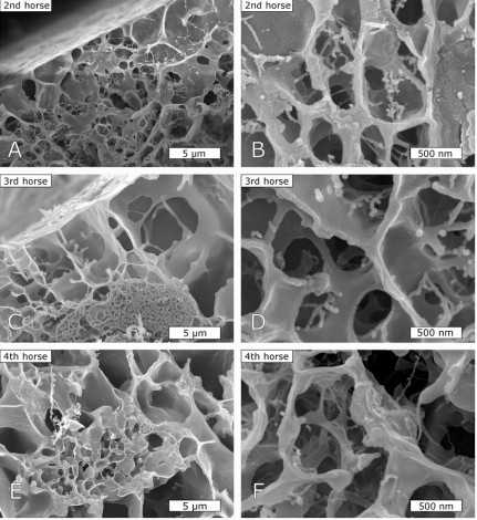
当在显微镜下观察时,肺粘膜看起来非常像海绵,将充满液体凝胶的大孔隔离开。Lehr教授说,“这些支架蛋白被称作粘蛋白(mucin)。”如今,研究人员证实纳米颗粒被困在这些结构之中,就像是被困在笼中一样。这就能解释为什么之前的很多研究发现粘膜中的纳米颗粒是高度移动性的,原因在于当前的研究是在纳米水平上开展的。在大孔内,这些纳米颗粒能够完全不受阻止地来回移动,因此,只有当他们试图越过当个孔时,它们受到阻挡而不能够逃离出去。
在这项研究中,研究人员使用了光镊子(optical tweezer)来证实了他们的发现:成束的激光被用来抓住和移动这些最小的颗粒。另一名通讯作者 Christian Wagner教授解释道,“我们能够使用光镊子的激光束来测量用来移动凝胶内颗粒所需的力量。我们能够以一种固定的力量在孔内的液体中移动颗粒。”
然而,无论何时颗粒达到孔壁时,激光束就是不能将它移到孔外。利用原子力显微镜和其他测试进行的研究进一步支持了这些发现:同样地,铁纳米颗粒能够穿过正常的对照凝胶,但是在磁铁作用下,它们也不能没有任何困难地穿过肺粘膜。利用低温电子显微镜,研究人员还对肺粘膜进行了结构分析。
他们期望对肺粘膜特殊结构的深入认识将有助于人们开发出新一代的药物来治疗呼吸道疾病。

In this study, the mobility of nanoparticles in mucus and similar hydrogels as model systems was assessed to elucidate the link between microscopic diffusion behavior and macroscopic penetration of such gels. Differences in particle adhesion to mucus components were strongly dependent on particle coating. Particles coated with 2 kDa PEG exhibited a decreased adhesion to mucus components, whereas chitosan strongly increased the adhesion. Despite such mucoinert properties of PEG, magnetic nanoparticles of both coatings did not penetrate through native respiratory mucus, resisting high magnetic forces (even for several hours). However, model hydrogels were, indeed, penetrated by both particles in dependency of particle coating, obeying the theory of particle mobility in an external force field. Comparison of penetration data with cryogenic scanning EM images of mucus and the applied model systems suggested particularly high rigidity of the mucin scaffold and a broad pore size distribution in mucus as reasons for the observed particle immobilization. Active probing of the rigidity of mucus and model gels with optical tweezers was used in this context to confirm such properties of mucus on the microscale, thus presenting the missing link between micro- and macroscopical observations. Because of high heterogeneity in the size of the voids and pores in mucus, on small scales, particle mobility will depend on adhesive or inert properties. However, particle translocation over distances larger than a few micrometers is restricted by highly rigid structures within the mucus mesh.
本网站所有内容来源注明为“梅斯医学”或“MedSci原创”的文字、图片和音视频资料,版权均属于梅斯医学所有。非经授权,任何媒体、网站或个人不得转载,授权转载时须注明来源为“梅斯医学”。其它来源的文章系转载文章,或“梅斯号”自媒体发布的文章,仅系出于传递更多信息之目的,本站仅负责审核内容合规,其内容不代表本站立场,本站不负责内容的准确性和版权。如果存在侵权、或不希望被转载的媒体或个人可与我们联系,我们将立即进行删除处理。
在此留言


#PNAS#
57
#肺粘液#
68
#纳米颗粒#
84