首次证实HCV也能感染血脑屏障的内皮细胞
2012-01-20 MedSci MedSci原创
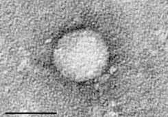
从细胞培养物中纯化出来的HCV病毒的电子显微镜图片,图片来自维基共享资源。 英国伯明翰大学科学家首次证实人大脑血脑屏障的内皮细胞能够被丙肝病毒(Hepatitis C virus, HCV)感染。 一个由病毒学家组成的研究小组发现大脑中内皮细胞拥有四个主要的蛋白受体,它们是HCV靶向血脑屏障(blood-brain barrier)所必需的。 2012年1月18日,这些发现作为研究亮点(

从细胞培养物中纯化出来的HCV病毒的电子显微镜图片,图片来自维基共享资源。
英国伯明翰大学科学家首次证实人大脑血脑屏障的内皮细胞能够被丙肝病毒(Hepatitis C virus, HCV)感染。
一个由病毒学家组成的研究小组发现大脑中内皮细胞拥有四个主要的蛋白受体,它们是HCV靶向血脑屏障(blood-brain barrier)所必需的。
2012年1月18日,这些发现作为研究亮点(Research Highlights)在线发表在Gastroenterology期刊上,表明除了肝脏肝细胞之外也有细胞对HCV感染比较敏感。
伯明翰大学免疫与感染学院Nicola Fletcher博士领导的研究人员与美国纽约市曼哈顿脑库(Manhattan Brain Bank)合作,检测了10名感染HCV的病人---他们死后捐献了大脑和肝脏组织---中4名的大脑HCV基因组物质,接着在实验室中证实从血脑屏障分离出的大脑细胞能够被HCV感染。
该论文通讯作者Jane McKeating教授说,“这是第一次报道中枢神经系统的细胞支持HCV复制。鉴于抗病毒治疗期间该病毒贮存库一直存在,这些观察可能用于临床应用。”
Fletcher解释道,“这些内皮细胞组成大脑的安全系统,类似于门边的保安,阻止不想要的东西进入。如果这种屏障遭受破坏,所有种类的物质能够进入大脑,这可能解释HCV感染的病人表现出的疲劳和其他症状。”
她说,因此保健治疗HCV感染的病人的当前标准只是部分有效的,人们有相当大的动力开发出靶向该病毒特异性酶的试剂作为替代性治疗方法,“我们期待相比于现存的药物,这样的试剂将更不可能跨越血脑屏障。我们相信我们的数据为传染性病原体如何能够靶向大脑提供详细的机制视图。”
HCV是一种黄病毒科(Flaviviridae) RNA病毒,造成全球的健康问题。HCV感染导致渐进性肝疾病,与包括中枢神经系统异常在内的多种肝外综合症(extrahepatic syndrome)相关联。(生物谷:towersimper编译)
Hepatitis C Virus Infects the Endothelial Cells of the Blood-Brain Barrier
Nicola F. Fletcher, Garrick K. Wilson, Jacinta Murray, Ke Hu, Andrew Lewis, Gary M. Reynolds, Zania Stamataki, Luke W. Meredith, Ian A. Rowe, Guangxiang Luo, Miguel A. Lopez–Ramirez, Thomas F. Baumert, Babette Weksler, Pierre–Olivier Couraud, Kwang Sik Kim, Ignacio A. Romero, Catherine Jopling, Susan Morgello, Peter Balfe, Jane A. McKeating
Background & Aims
Hepatitis C virus (HCV) infection leads to progressive liver disease and is associated with a variety of extrahepatic syndromes, including central nervous system (CNS) abnormalities. However, it is unclear whether such cognitive abnormalities are a function of systemic disease, impaired hepatic function, or virus infection of the CNS.
Methods
We measured levels of HCV RNA and expression of the viral entry receptor in brain tissue samples from 10 infected individuals (and 3 uninfected individuals, as controls) and human brain microvascular endothelial cells by using quantitative polymerase chain reaction and immunochemical and confocal imaging analyses. HCV pseudoparticles and cell culture–derived HCV were used to study the ability of endothelial cells to support viral entry and replication.
Results
Using quantitative polymerase chain reaction, we detected HCV RNA in brain tissue of infected individuals at significantly lower levels than in liver samples. Brain microvascular endothelia and brain endothelial cells expressed all of the recognized HCV entry receptors. Two independently derived brain endothelial cell lines, hCMEC/D3 and HBMEC, supported HCV entry and replication. These processes were inhibited by antibodies against the entry factors CD81, scavenger receptor BI, and claudin-1; by interferon; and by reagents that inhibit NS3 protease and NS5B polymerase. HCV infection promotes endothelial permeability and cellular apoptosis.
Conclusions
Human brain endothelial cells express functional receptors that support HCV entry and replication. Virus infection of the CNS might lead to HCV-associated neuropathologies.
本网站所有内容来源注明为“梅斯医学”或“MedSci原创”的文字、图片和音视频资料,版权均属于梅斯医学所有。非经授权,任何媒体、网站或个人不得转载,授权转载时须注明来源为“梅斯医学”。其它来源的文章系转载文章,或“梅斯号”自媒体发布的文章,仅系出于传递更多信息之目的,本站仅负责审核内容合规,其内容不代表本站立场,本站不负责内容的准确性和版权。如果存在侵权、或不希望被转载的媒体或个人可与我们联系,我们将立即进行删除处理。
在此留言


#首次证实#
57
#HCV#
78
#血脑屏障#
72