JACC:第三军医大学新桥医院报道心脏副神经节瘤一例
2013-05-07 JACC dxy
49岁女性患者,因不典型胸部不适伴活动后气短半年,到我院心外科就诊。体格检查、胸部X片以及实验室检查指标等均无显著发现,胸部CT血管成像发现中纵隔有一肿物。 经食管超声(TEE)发现该肿块为心包内异质性团块,位于左、右心房与主动脉根部区域,有一起自主动脉的血管供血,血供丰富。 实时三维(RT-3D)TEE进一步明确其位置在双心房-主动脉根部的间隙中,并对
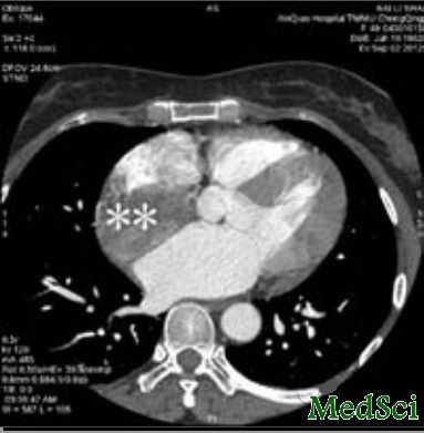
49岁女性患者,因不典型胸部不适伴活动后气短半年,到我院心外科就诊。体格检查、胸部X片以及实验室检查指标等均无显著发现,胸部CT血管成像发现中纵隔有一肿物。
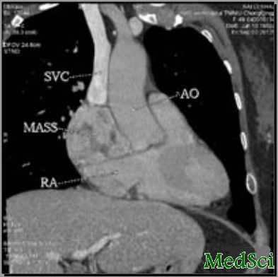
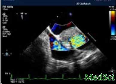
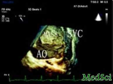
经食管超声(TEE)发现该肿块为心包内异质性团块,位于左、右心房与主动脉根部区域,有一起自主动脉的血管供血,血供丰富。
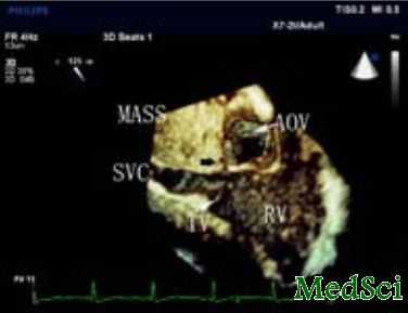
实时三维(RT-3D)TEE进一步明确其位置在双心房-主动脉根部的间隙中,并对上腔静脉造成压迫。
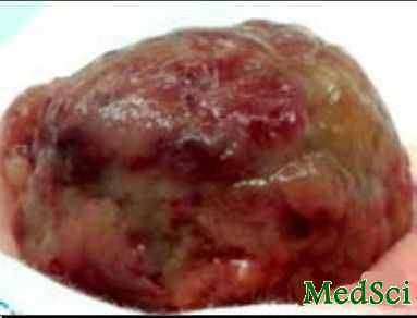
在TEE发现的基础上,我们判定其包膜完整,未侵犯周围组织。我们制定了体外循环下外科切除的手术计划,并成功进行。
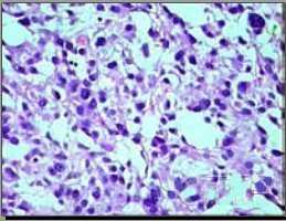
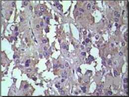
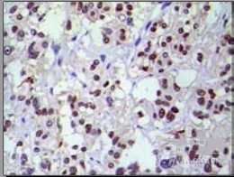
术后随访6月以上,患者恢复良好。病理回报,细胞形态相似,胞浆富含颗粒(高倍,40X; H&E染色)。免疫组化显示CgA与S-100(I,J 40 X)阳性,提示心脏副神经节瘤诊断无误。
该病十分罕见。可通过RT-3D-TEE可明确其位置以及与周围组织关系。
病例报道者:第三军医大学新桥医院超声科夏红梅。将发表在JACC(Journal of the American College of Cardiology),该杂志最新的影响因子为14.2,在心脏病领域中排名第二,仅次于circulation。
与心脏相关的拓展阅读:
Cardiac Paraganglioma
A 49-year-old female presented to the department of cardiovascular surgery with short breath after activity and atypical chest discomfort during the past half a year. Physical examination, initial chest X-ray, and laboratory values were unremarkable. Computed tomography angiogram of the chest disclosed a mass (**) in middle mediastina (A and B). Transesophageal echocardiography (TEE) revealed a 6-cm heterogeneous intrapericardial mass in the left atrium (LA)–right atrium (RA)–aortic root area with plentiful blood supply by a nourishing vessel originating from the aorta (AO) (C and D, Online Videos 1 and 2). Real-time 3-dimensional TEE further delineated the tumor location in the atria–aortic root window, which was compressing the superior vena cava (SVC) (E and F). Based on the TEE findings of an encapsulated tumor without surrounding invasion, surgical excision by extracorporeal circulation was planned and performed successfully (G). Recovery was achieved over a 6-month follow-up. Microscopic examination of the tissue biopsies showed sheaths of uniform cells with abundant granular cytoplasm (40×; H&E stain) (H). Later, immunohistochemical staining of the tissue was positive for CgA and S-100 (40×) (I and J), which confirmed the diagnosis of a paraganglioma. Cardiac paraganglioma is extremely rare. Real-time 3-dimensional TEE can provide detailed information in diagnosing cardiac paraganglioma location and relation to neighbor. AOV = aortic valve; RV = right ventricle; TV = tricuspid valve.
本网站所有内容来源注明为“梅斯医学”或“MedSci原创”的文字、图片和音视频资料,版权均属于梅斯医学所有。非经授权,任何媒体、网站或个人不得转载,授权转载时须注明来源为“梅斯医学”。其它来源的文章系转载文章,或“梅斯号”自媒体发布的文章,仅系出于传递更多信息之目的,本站仅负责审核内容合规,其内容不代表本站立场,本站不负责内容的准确性和版权。如果存在侵权、或不希望被转载的媒体或个人可与我们联系,我们将立即进行删除处理。
在此留言





#JACC#
55
#军医#
47
#ACC#
49
#副神经节瘤#
64